Common Dental Myths
By Dr. Minal Patil
B.D.S, M.D.S. (Periodontist And Implantologist)
Chest Tightness: Causes, Symptoms, and Home Remedies
What is Chest Tightness?
Chest tightness is a feeling of pressure, heaviness, fullness, or discomfort in the chest. Some people describe it as a weight pressing on the chest or difficulty taking a deep breath.
Although chest tightness is often associated with a heart attack, many cases are caused by non-cardiac conditions such as respiratory problems, acid reflux, anxiety, or muscle strain. It may occur suddenly or develop gradually depending on the underlying cause.
Causes of Chest Tightness
Chest tightness can occur due to several medical conditions, including:
- Asthma
- Pneumonia
- Chronic Obstructive Pulmonary Disease (COPD)
- Pulmonary Embolism
- Respiratory Infections
- Pulmonary Hypertension
- Allergies
- Acid Reflux (GERD)
- Coronary Artery Disease (CAD)
- Mitral Valve Prolapse
- Pericarditis
- Gallstones
- Shingles
- Peptic Ulcer
- Anxiety or Panic Attacks
Symptoms of Chest Tightness
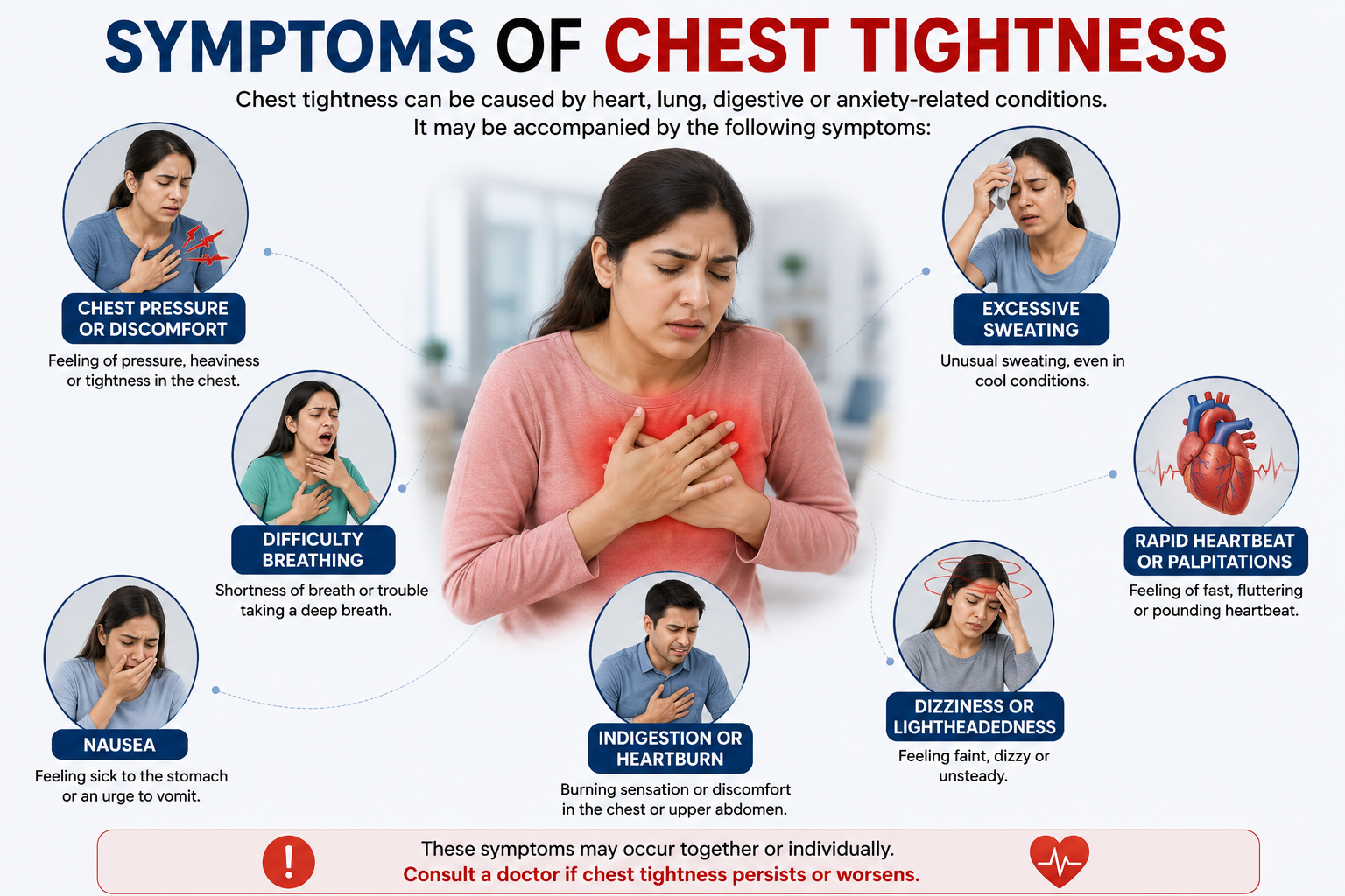
Chest tightness may be accompanied by:
- Chest pressure or discomfort
- Difficulty breathing
- Nausea
- Indigestion
- Excessive sweating
- Dizziness or lightheadedness
- Rapid heartbeat or palpitations
Risk Factors
The following factors may increase the risk of chest tightness:
- Obesity or overweight
- Family history of heart or lung disease
- Smoking
- Allergy exposure
- Sedentary lifestyle
Home Remedies for Chest Tightness
Depending on the underlying cause, the following measures may help relieve chest tightness:
Respiratory Problems
- Drink plenty of fluids.
- Use a humidifier.
- Try steam inhalation or vapour rubs.
- Take prescribed decongestant medications if advised by your doctor.
Gastrointestinal Problems
- Eat smaller, frequent meals.
- Avoid smoking.
- Avoid spicy, fatty, caffeinated, and carbonated foods and drinks.
Lung Conditions
- Get adequate rest.
- Avoid known triggers.
- Take medications as prescribed.
Anxiety or Panic Attacks
- Practice deep breathing exercises.
- Try meditation or mindfulness techniques.
- Exercise regularly to reduce stress.
Muscular Injuries
- Rest the affected muscles.
- Apply ice packs if recommended.
- Seek medical advice for persistent pain.
When Should You Consult a Doctor?
Seek immediate medical attention if chest tightness is accompanied by:
- Chest pain spreading to the neck, jaw, or left arm
- Persistent shortness of breath
- Severe dizziness or fainting
You should also consult a doctor if you experience:
- Fever
- Unexplained weight loss
- Rapid heartbeat or palpitations
- A history of asthma, COPD, or other lung diseases
- Persistent or worsening chest tightness
Conclusion
Chest tightness can occur due to a variety of heart, lung, digestive, or anxiety-related conditions. While some cases are mild and improve with simple home remedies, persistent or severe chest tightness should never be ignored. Early medical evaluation helps identify the underlying cause and ensures timely treatment, reducing the risk of serious complications.
Gum Disease
By Dr. Minal Patil
(Periodontist / Gum Specialist)
Dangerous Heart Rate vs Normal Heart Rate: Know the Difference
Table of Content
What is the Normal Heart Rate?
What is a Dangerous Heart Rate?
When is the Heart Rate Too High?
When is the Heart Rate Too Low?
When to Contact Healthcare?
Conclusion
What is the Normal Heart Rate?
When an individual is at rest, their heart pumps the least amount of blood necessary to sustain their body, which is known as the resting or normal heart rate of the human body. A normal heartbeat range is typically between 60 to 100 beats per minute (BPM) at rest for adults. However, some individuals may have a resting heart rate that is below 60 beats per minute and be regarded as normal.
Factors like age, fitness level, and medication can influence this range. For instance, athletes may experience a drop in heart rate—up to 40 beats at times. In addition, people who take certain medications, such as beta-blockers, could also have a reduced heart rate at rest.
What is a Dangerous Heart Rate?
A dangerous heart rate occurs when the pulse deviates significantly from the standard range. The term “tachycardia” refers to an excessively high heart beat (over 100 beats per minute). However, an individual’s age and general health may also play a pivotal role in determining what is considered to be too fast.
Conversely, bradycardia is when the heart rate drops below 60 BPM, signalling a potentially risky condition. A heart rate of less than 60 beats per minute is common, even healthy, for athletes and frequent exercisers. Treatment may not be necessary for borderline or occasional bradycardia. However, if bradycardia is left untreated for an extended period of time, it might worsen. It may cause fainting and dizziness.
When is the Heart Rate Too High?
A high heart rate, especially persisting over extended periods, might indicate underlying health issues. High heart rates can result from stress, intense physical activity, dehydration, or underlying conditions like hyperthyroidism. Consistently elevated rates might lead to dizziness, chest pain, or fainting, necessitating immediate attention.
The following are factors that might cause high heart rate:
Fever
Overindulgence in drinking or alcohol withdrawal
Elevated amounts of caffeine
Blood pressure, high or low
Imbalance of electrolyte elements, which include potassium, sodium, calcium, and magnesium, in the blood
Adverse effects of medication
Use of illicit substances, such as methamphetamine or cocaine, which are stimulants.
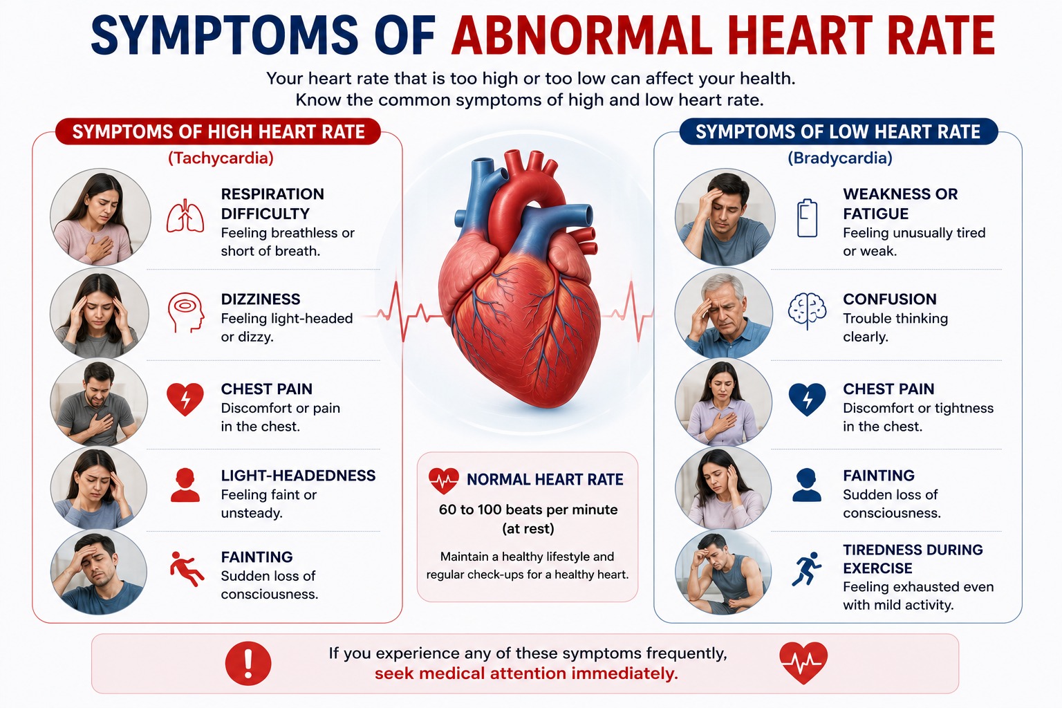
Symptoms of High Heart Rate
Respiration difficulty
Dizziness
Chest Pain
Light-headedness
Fainting
When is the Heart Rate Too Low?
A low heart rate could be a sign of a well-conditioned heart, but it could also indicate health concerns. Factors contributing to a slow heart rate include ageing, medications, heart tissue damage, electrolyte imbalances, or conditions affecting the heart’s electrical system.
Low heart rate can be caused by:
A damage to the heart tissue brought on by ageing
A birth defect involving the heart
Inflammatory conditions like lupus or rheumatic fever
A cardiac surgery
An imbalance of blood components like calcium or potassium
Sleeping with frequent pauses in breathing
The brain may not receive enough blood flow due to a dangerously low heart rate.
Signs of Low Heart Rate
Weakness or fatigue
Confusion
Fainting
Chest pain
Feeling extremely tired when exercising
A dangerously low heart rate requires prompt medical management.
When to Contact Healthcare?
In certain cases, fluctuations in heart rate could signify potential dangers. Seek medical advice if experiencing persistent irregular heartbeats, chest pain, shortness of breath, or if the heart rate consistently exceeds or falls below the heart rate normal range.
Moreover, if symptoms persist despite rest, it’s crucial to consult a healthcare professional promptly. Individuals with existing heart conditions, irregular heartbeats, or other health conditions should regularly monitor their heart rate and consult healthcare providers for any alarming changes.
Conclusion
Understanding the delicate balance between a normal heart rate range and a dangerous heart rate range is essential for maintaining optimal health. While slight fluctuations can be normal, persistently high or low heart rates warrant attention.
A resting heart rate is a vital sign of several underlying problems as well as heart health in general. Thus, it should be constantly monitored. Regular check-ups, a healthy lifestyle, and prompt medical attention for concerning symptoms are vital in ensuring a healthy heart rate and overall well-being.
Everyone must always prioritize monitoring and addressing any irregularities in the heart rate to safeguard their well-being.
Gum Disease
By Dr. Minal Patil
(Periodontist / Gum Specialist)
Left Ventricular Dysfunction (LV Dysfunction): Symptoms, Causes, Diagnosis and Treatment
Table of Content
- What is Left Ventricular Dysfunction (LV Dysfunction)?
- Symptoms of Left Ventricular Dysfunction
- Causes of Left Ventricular Dysfunction
- Tests to Diagnose Left Ventricular Dysfunction
- Left Ventricular Dysfunction Treatment
- How can I prevent left-sided heart failure?
- Management of LVD with Lifestyle Changes
- Changes in Heart with LVD
- Conclusion
- FAQs
The heart serves as the body’s central pumping station, ensuring oxygen-rich blood which is efficiently distributed throughout the body. The left ventricle, one of the heart’s four chambers, plays an essential role in this process. The left ventricle’s primary job is to pump blood rich in oxygen to every region of the body. When the left ventricle is unable to function properly, it can lead to a medical condition known as Left Ventricular Dysfunction (LV Dysfunction). This medical condition of the heart can have a significant impact on a person’s health and overall well-being.
What is Left Ventricular Dysfunction (LV Dysfunction)?
Left Ventricular Dysfunction, often referred to simply as LV Dysfunction, is a condition in which the left ventricle of the heart does not contract as effectively as it should. This reduced ability to contract and pump blood is commonly associated with a condition called heart failure, which can be either systolic heart failure or diastolic heart failure. LV Dysfunction can be further classified into two main types:
- Systolic Heart Failure – Left ventricular systolic dysfunction causes the ventricle to have trouble contracting firmly, which lowers the ejection fraction, the amount of blood the ventricle pumps out with each beat. Around 55-70% is considered a standard ejection. Systolic dysfunction, which reduces the heart’s capacity to pump blood efficiently, is indicated when it drops below this range.
- Diastolic Heart Failure – The inability of the left ventricle to properly relax and fill during the diastolic portion of the cardiac cycle is known as diastolic dysfunction. This causes the left ventricle’s pressure to rise and its ventricular filling to be compromised.
Symptoms of Left Ventricular Dysfunction
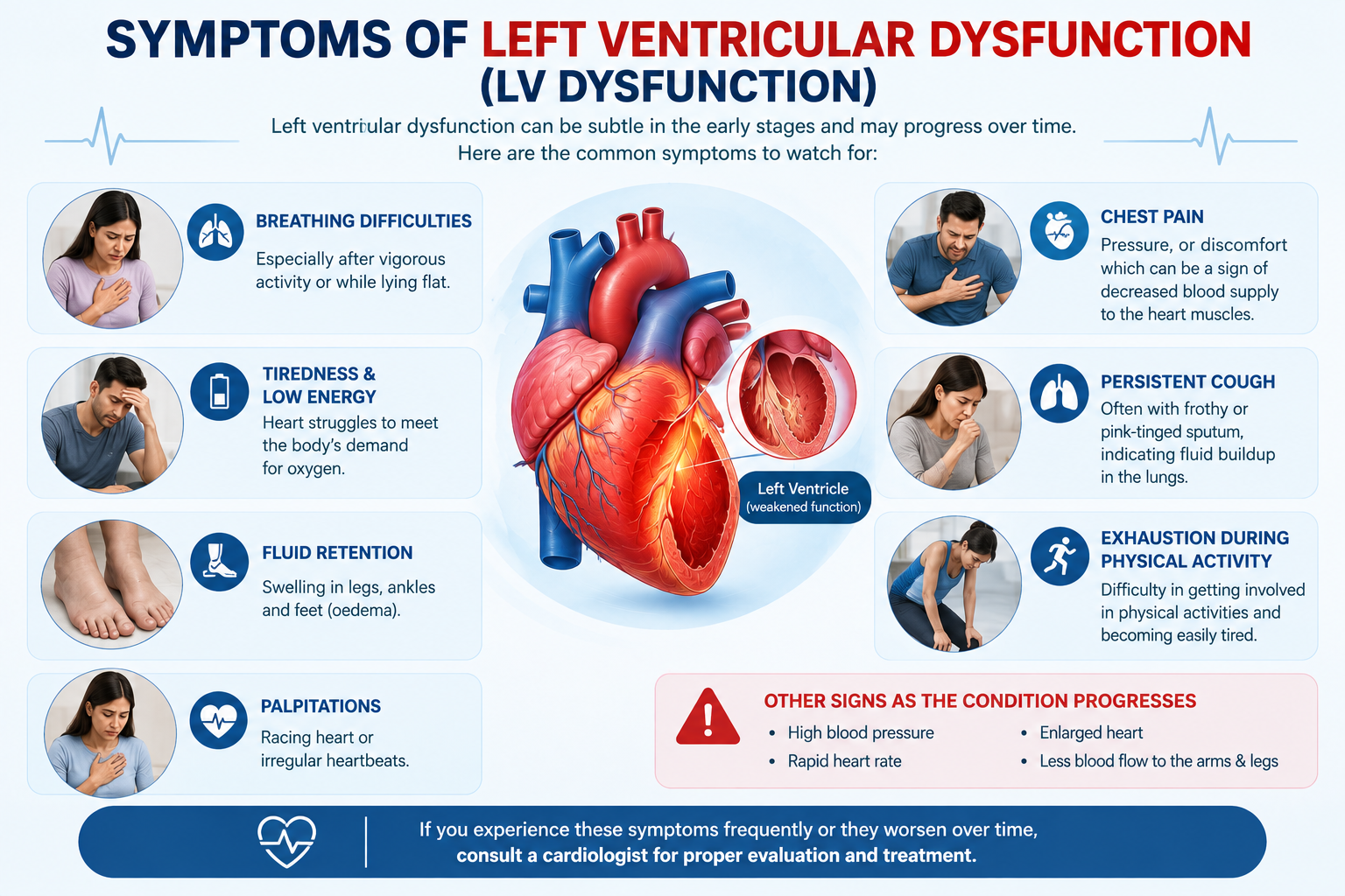
Left ventricular dysfunction symptoms can be subtle in the early stages and may progress over time. Common symptoms of severe left ventricular dysfunction include:
- Breathing difficulties are a typical sign of left ventricular dysfunction, particularly after vigorous activity or while the patient is lying flat.
- Persistent tiredness and a lack of energy can be early signs of left ventricular dysfunction, as the heart struggles to meet the body’s demands for oxygen.
- Fluid retention can cause swelling in the legs, ankles, and feet, a condition known as oedema. This occurs because the heart’s inability to pump blood effectively can cause fluid buildup in the body.
- Individuals with left ventricular dysfunction may experience palpitations, a racing heart, or irregular heartbeats.
- Some patients may suffer chest pain, pressure, or discomfort, which can be the symptoms of decreased blood supply to the heart muscles.
- A persistent cough, typically accompanied by frothy or pink-tinged sputum, might be an indication of fluid buildup in the lungs.
- Those suffering from LV dysfunction may find it difficult to get involved in physical activities and may become easily exhausted during exercise.
As time passes, the heart may attempt to compensate by pounding with greater force. This leads to damage, which may result in the following:
- High blood pressure
- Rapid heart rate
- Enlarged heart
- Less blood flow to the arms & legs
Causes of Left Ventricular Dysfunction
Left ventricular dysfunction can result from a variety of underlying causes. Some common causes of LV dysfunction include:
- Coronary Artery Disease (CAD) – The most common cause of LV dysfunction is CAD, which occurs when the coronary arteries supplying the heart muscle become narrowed/blocked by a buildup of cholesterol and plaque.
- Hypertension (High Blood Pressure) – Prolonged high blood pressure can lead to LV dysfunction by causing the left ventricle of the heart to work harder to pump blood, leading to thickening and stiffening of the heart muscle.
- Valvular Heart Disease – Heart valve problems can cause abnormalities in the heart’s blood flow and pressure, which may result in LV dysfunction.
- Cardiomyopathy – Cardiomyopathy is a group of heart muscle diseases that can weaken the heart and lead to LV dysfunction.
- Diabetes – Blood arteries may harden as a result of high blood sugar. It can lead to muscle thickening since it puts more strain on the heart.
- Obesity – Being overweight or obese increases the strain on the heart and can contribute to LV dysfunction.
- Obstructive Sleep Apnea (OSA) – A patient with OSA will cease breathing while asleep. The body undergoes a complicated series of modifications as a result. These modifications include elevated blood pressure, decreased cardiac oxygen supply, and heightened nervous system activity. The supply and demand of oxygen are not matching as a result of these changes. It further increases the chance of developing additional cardiac diseases, including diastolic and systolic heart failure.
- Kidney Disease – Chronic kidney disease might lead to the condition of fluid retention and increased strain on the heart.
- Family History – A family history of heart disease may increase the risk of developing LV Dysfunction.
- Age – As individuals age, the risk of LV dysfunction increases as the heart undergoes natural changes.
- Sedentary lifestyle – Anybody who is physically inactive runs an increased risk of obesity, diabetes, coronary heart disease, and high blood pressure. Any of these disorders can cause diastolic heart failure or LV dysfunction.
Tests to Diagnose Left Ventricular Dysfunction
Several tests and procedures are commonly used to confirm the presence of LV dysfunction and determine its underlying cause. These diagnostic tools may include:
- Echocardiogram – An echocardiogram, or cardiac ultrasound, uses sound waves to create detailed images of the heart’s structure and function. It can assess the size of the heart chambers, ejection fraction, and the presence of any valve abnormalities.
- Electrocardiogram (ECG or EKG) – An ECG records the electrical activity of the heart, helping to identify any abnormal heart rhythms or patterns.
- Cardiac MRI (Magnetic Resonance Imaging) – A cardiac MRI provides detailed images of the heart, allowing for a more in-depth assessment of cardiac structure and function.
- Lab Test – Tests on the blood and urine might be performed to look for conditions that could compromise heart health. Tests may be performed in order to evaluate liver & kidney function, blood sugar, and cholesterol levels.
- Cardiac Catheterisation – In this invasive procedure, a thin, flexible tube (catheter) is inserted into the heart’s blood vessels to measure pressure, take samples, and assess blood flow.
- Stress Tests – Stress tests, including exercise stress tests or pharmacological stress tests, can evaluate how the heart responds to increased workload and detect any areas of reduced blood flow.
Left Ventricular Dysfunction Treatment
Left ventricular dysfunction treatment can involve a combination of lifestyle modifications, medications, and surgeries for severe left ventricular dysfunction treatment:
Lifestyle Modifications:
- A heart-healthy diet that is low in salt and saturated fats can help manage LV dysfunction. Reducing sodium intake is crucial to prevent fluid retention.
- Regular physical activity, as per the guidance of a healthcare provider, can improve heart health and overall well-being.
- The stress on the heart can be lessened by achieving and maintaining a healthy weight.
- Quitting smoking is essential for cardiovascular health.
- Addressing substance abuse and excessive alcohol consumption is crucial.
Medications:
- Diuretics help reduce fluid retention by promoting the excretion of excess fluids through the kidneys.
- ACE Inhibitors or ARBs can help relax blood vessels and lower blood pressure.
- Beta-blockers lessen the burden on the heart by slowing the heart rate.
- Calcium Channel Blockers can help relax blood vessels and lower blood pressure.
- Digoxin may be used to improve heart function and control heart rate.
- Adversary of the mineralocorticoid receptor eliminates extra fluid and salt from the body while retaining potassium.
- To decrease cholesterol levels and lessen the risk of atherosclerosis, doctors may prescribe statins.
Surgery:
- Angioplasty & Stent Placement – In cases of coronary artery disease, angioplasty and stent placement may be performed to open blocked or narrowed arteries.
- Coronary Artery Bypass Grafting (CABG) – CABG surgery can bypass blocked coronary arteries to improve blood flow to the heart.
- Valve Repair or Replacement – If valvular heart disease is contributing to LV dysfunction, surgical repair or replacement of heart valves may be necessary.
- Implantable Devices – Some patients may benefit from implantable devices like pacemakers or defibrillators to regulate heart rhythms.
- Heart Transplant – In severe cases of LV dysfunction, a heart transplant may be considered when other treatment options are no longer effective.
Conclusion
Left Ventricular Dysfunction is a complex and potentially life-threatening condition that can significantly impact an individual’s quality of life. Early detection and effective management are crucial in providing the best outcomes for those affected by this medical condition. As with many cardiovascular conditions, prevention is key, and individuals can take specific steps to reduce their risk of developing LV dysfunction by maintaining a healthy lifestyle.